
Med Lightbox
Multi-user medical image viewer and annotation tool
Requires MouseMux running on Windows. Get it free.
Med Lightbox
Multi-user medical image viewer and annotation tool.
What This Is
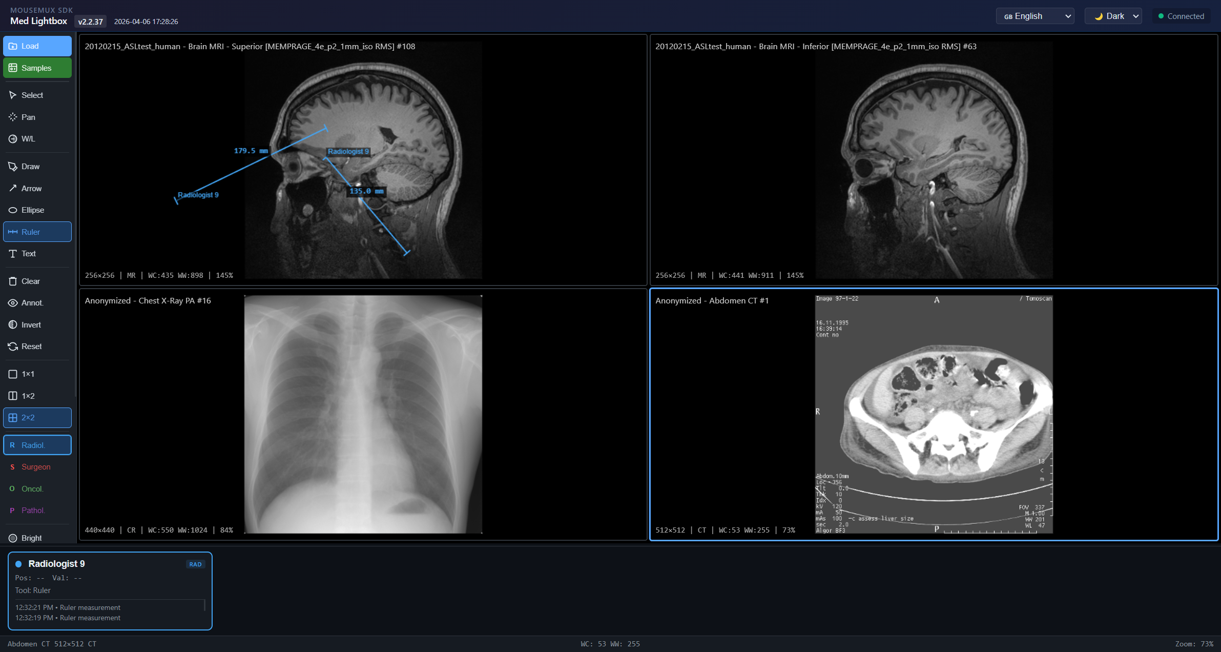
A collaborative medical image viewer where multiple clinicians can simultaneously view, annotate, and measure medical images in real-time. It supports DICOM files (X-ray, CT, MRI) and standard image formats (PNG, JPG, BMP, TIFF, WebP). Each user gets their own cursor, tools, and role assignment, so a radiologist, surgeon, and oncologist can all review the same images together, each marking up what they see with their own color-coded annotations. Built with the MouseMux Web SDK for multi-user input.
How It Works
The interface has a viewport grid in the center, a toolbar on the left, and per-user status panels at the bottom. The viewport grid can show 1, 2, or 4 images at once in a 1x1, 1x2, or 2x2 layout. Each viewport independently displays its own image with separate zoom, pan, and window/level settings. You can load images through a file picker or drag-and-drop onto specific viewports. Demo DICOM images load automatically on startup to get you going.
The annotation tools include freehand drawing, arrows, ellipse ROI markers, ruler measurements, and text labels. Rulers show real distances in millimeters when the DICOM file includes pixel spacing data. Ellipses show area in mm or cm squared. All annotations are stored in image coordinates so they stay in place when you zoom and pan. You can select, move, and delete any annotation with the pointer tool.
Each user picks a clinical role: Radiologist, Surgeon, Oncologist, or Pathologist: which sets their annotation color and shows a role badge on their status panel. There’s also a set of image processing algorithm overlays including threshold detection, edge detection, heat maps, tissue segmentation, CLAHE contrast enhancement, and more. Each user can independently toggle these overlays without affecting what others see. The window/level tool adjusts brightness and contrast by dragging: horizontal for width, vertical for center: which is the standard approach for reading medical images.
Getting Started
Launch the app from the MouseMux launcher and connect when prompted. The app loads demo DICOM images automatically in a 2x2 grid. Pick your clinical role from the toolbar buttons (R for Radiologist, S for Surgeon, etc.) and select an annotation tool to start marking up images. Use the scroll wheel to zoom and right-click drag to pan. The W/L tool adjusts windowing for DICOM images. You can load your own DICOM or standard image files through the file picker or by dragging them onto a viewport.
MouseMux Development Team · https://www.mousemux.com